
Wart or Mimic?
Case 1: During a routine examination, an asymptomatic wart like lesion is noted near the axilla of a 35-year-old woman.
The patient says that it has been present since childhood. Is this a wart . . . or something else?
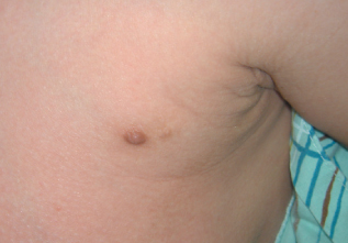
Case 1: This is an accessory nipple, which can occur along the milk line in both men and women. Unlike warts, these growths are soft and sometimes have a central dell.
Case 2: A 70-year-old wheelchair-bound man who has had one leg amputated and lives in a skilled nursing facility complains of a “wart” on his amputation stump. Does this look like a wart to you?
Case 2: This patient has amputa- tion stump hyperkeratosis, which is thought to be caused by the per- sistent rubbing that results from an ill-fitting prosthesis. These lesions are larger than warts and do not display the black “dots” that are characteristic of warts with throm- bosed capillaries. Moreover, no human papillomavirus is found on polymerase chain reaction testing. Sometimes the keratotic tops of amputation stump hyperkeratoses can be peeled away like those of actinic keratoses.
Case 3: These are calluses, which can be distinguished from warts in 3 ways:
•Calluses do not contain the throm- bosed capillaries (black dots) that are characteristic of warts.
•They do not interrupt the normal skin markings (dermatoglyphs), as warts do.
•Calluses are more painful on di- rect than on lateral pressure, while warts are more painful on lateral than on direct pressure.
Case 4: This lesion has bothered a healthy 16-year-old boy. Can you identify it?
Case 4: This is a common wart. The wart is keratotic and displays the characteristic interrupted skin lines (dermatoglyphs).
Case 5: This is a filiform wart. These warts have fingerlike projections whose ends can be irregular or brushlike. Filiform warts resemble skin tags, but the latter are soft
Case 6: These rough, irregular papules arose under the nail of a healthy 16-year-old boy. What does this look like to you?

This is a subungual/peri- ungual wart, a keratotic papule that resists treatment. Subungual warts underlie and infiltrate the nail fold; periungual warts abut the nail fold. These warts are more difficult to eradicate than other types of warts because a nidus of infected tissue remains after chemical or surgical treatment, which allows for reexten- sion of the wart.
Case 7: What type of outbreak is seen here on the face of a healthy 33-year-old man?
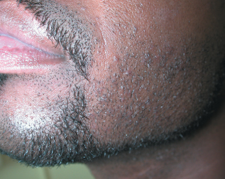
Case 7: These are flat warts, slight- ly elevated, flat-topped papules that are usually smaller than 5 mm. They typically occur in shaved areas, such as the beard area in men and lower legs in women. Shaving may spread the outbreak. Flat warts are pink
in pale-skinned persons and brown in darker-skinned persons
Case 8: What type of lesion is seen here in a healthy 30-year-old woman? The patient says that the hyperpigmented area has been present since birth.
This is a congenital nevus. These lesions are softer than warts and sometimes cerebriform, although they can also be verrucous. However, they have no black dots. In the groin, congenital nevi may be mistaken for condy- lomata acuminata, seborrheic keratoses, or skin tags; a biopsy is sometimes required to confirm the diagnosis.