
Peer Reviewed
Dermal Eccrine Cylindroma
Author:
Joe R. Monroe, MPAS, PA
Epiphany Dermatology, Tulsa, Oklahoma
Citation:
Monroe JR. Dermal eccrine cylindroma [published online February 11, 2019]. Consultant360.
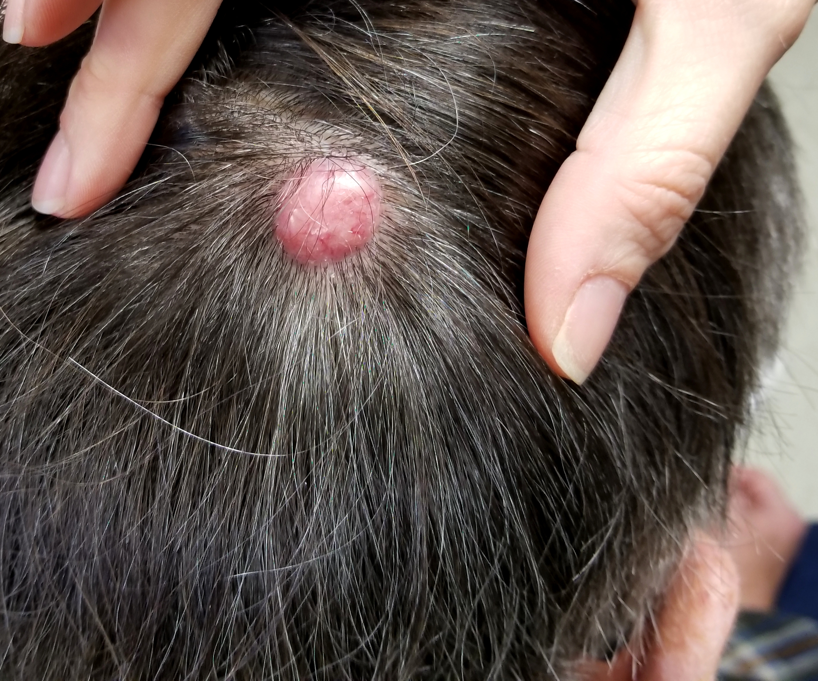
A 50-year-old man presented with concern for a scalp lesion that had been present for most of his life. It had caused few if any problems until lately, when it had begun to enlarge, causing it to be caught often with his comb and his fingernails.
He denied having been born with the lesion, but he could remember having it as a small child.
Physical examination. The lesion was in the left parietal scalp, measuring almost 2 cm (Figure). It was a smooth, firm, round, pink nodule, the surface of which was somewhat telangiectatic. It was attached to the scalp by a 1 cm pedicle. The patient’s skin was not especially sun-damaged, and he had never had skin cancer.
The lesion was excised with 4-mm margins, down to adipose tissue, with primary closure. The specimen was sent for pathologic examination, the results of which revealed islands of adnexal cells with hyperchromatic nuclei, which formed palisades focally. No inflammatory infiltrate was present, a finding that served to distinguish this lesion from other possibilities such as spiradenoma. The deep margins were clear.
Discussion. The correct diagnosis in this patient’s case was dermal eccrine cylindroma, a rare benign adnexal lesion. A cylindroma is so named because of the nests of cells seen on pathologic cross-sections that resemble cylinders.
These benign tumors can be solitary or multiple and often coalesce into turban-like scalp and/or forehead lesions. Aside from being unsightly, they have no other ominous implications.
In terms of a differential diagnosis, solitary lesions such as this one can closely resemble basal cell carcinoma or even neurofibroma.
The only treatment is surgical, which can become quite extensive with larger lesions.