
Peer Reviewed
What Is the Cause of an Older Patient’s Persistent Hip Pain?
Introduction. A 63-year-old man presented to his primary care physician’s clinic with left hip pain after sustaining a fall while roller skating 2 years earlier.
History. The patient had no significant past medical history. He described his pain as a constant throbbing that radiates down the anterior aspect of his left thigh to his knee. He endorsed soreness but denied edema, cramping, or paresthesias. He rated his pain at the time of the fall as a six out of 10.
During the 2 years following his fall, the patient saw four clinicians. He was experiencing worsening pain, which caused him to stop roller skating. He then developed left hip catching, where he would feel a popping, snapping, or hear a clicking sound during movement. The patient denied any alleviating factors.
At the time of presentation, the patient reported worsening pain, rated at 10 out of 10 in intensity, radiating down to his left foot. On physical examination, his hip demonstrated full range of motion, and the pain was not reproduced with log roll or resisted flexion. Neurologic examination revealed intact sensation and preserved strength (5/5) without pelvic laxity or overlying erythema. However, he exhibited marked tenderness to palpation over the left greater trochanter and sacroiliac joint. Provocative maneuvers, including flexion adduction internal rotation and flexion abduction external rotation, were positive, as was the straight leg raise test.
Diagnostic Testing. The evaluating clinicians obtained AP (Figure 1) and lateral (Figure 2) radiographs as well as magnetic resonance arthrogram (MRA) images of the left hip (Figures 3 and 4).
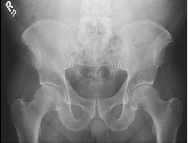
Figure 1. Anterior posterior (AP) radiograph of the pelvis is shown.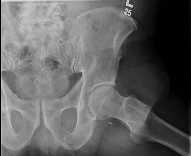
Figure 2. A lateral radiograph of the left hip is shown.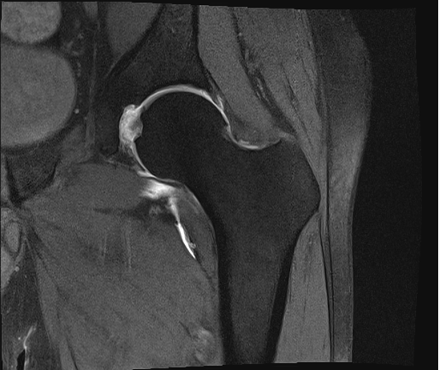
Figure 3. The Coronal T2 MRI with arthrogram is shown.
Figure 4. Axial T2 MRI of the hip with arthrogram is shown.