
Peer Reviewed
Can You Diagnose the Lesions on This 21-Year-Old Woman’s Lower Extremities After She Returned from the Philippines?
Introductory sentence. A 21-year-old previously healthy woman presented to the emergency room with a progressive rash on her right leg and was found to be febrile and hypotensive.
History. The woman presented to the emergency room following 1 day of fatigue and the development of a progressive rash after returning from a trip to the Philippines. The patient reported that the lesions first appeared on her right lower extremity while she was in the Philippines visiting her family for 1 month. During that time, she visited the Island of Boracay, where she swam in both fresh and seawater, traveled to Bataan, and drank iced tea from a roadside vendor after hiking a mountain. She also had exposure to many flies and incurred bites from them. The patient did not visit a clinician at this time while in the Philippines.
When she returned to the United States, the lesions increased in number and became tender with palpation or standing (Figure 1). The lesions spread to her left lower extremity and finally reached her lower back (Figure 2). She noted that pus drained from a few of them after manipulation. The patient reported no previous sexual activity and is up to date on all her immunizations. She does not take any daily medications besides a multivitamin. Review of systems was negative for all symptoms other than a single day of fatigue and presence of a rash.
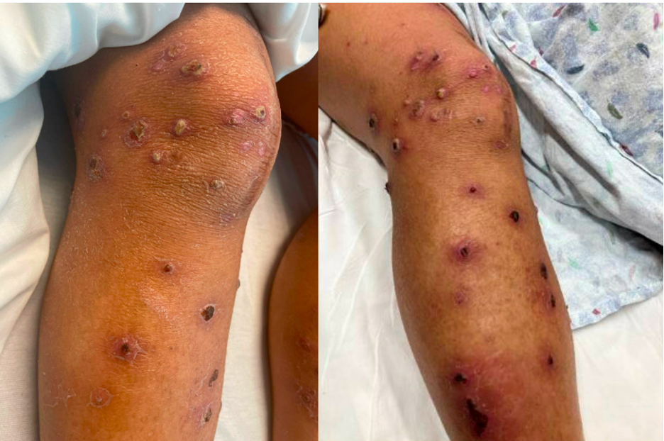
Figure 1. Discrete keratotic, pustular papulonodular lesions with central necrotic crust in differing stages of healing on the lower extremities.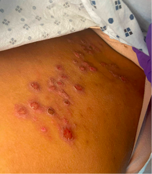
Figure 2. Discrete papulonodular lesions with superficial erosions in a grouped cluster on the lower back and upper buttocks.Diagnostic testing. In the emergency room, the patient had a fever of 102.6℉, tachycardia of 148 bpm, blood pressure of 109/48, and leukocytosis of 24.06 K/uL (88% neutrophils). The patient was well-appearing and in no acute distress.
Her skin examination demonstrated papulonodular lesions with superficial erosions in a grouped cluster on the lower back and upper buttocks as well as discrete keratotic, pustular papulonodular lesions with central necrotic crust in differing stages of healing on the lower extremities. Other notable laboratory values included normal chemistry panel, liver function test, negative antinuclear antibodies (ANA), anti-neutrophil cytoplasmic antibodies (ANCA), methicillin-resistant Staphylococcus aureus (MRSA), rapid plasma reagin (RPR), human immunodeficiency (HIV) antibodies, hepatitis B antigen, and hepatitis C antibody tests. Blood cultures were negative at 24 hours. A swab of a superficially ulcerated lesion was negative for Herpes Simplex Virus (HSV) and Varicella Zoster Virus (VZV).
Dermatology was consulted and performed a deep shave biopsy of characteristic lesions for tissue culture and histopathologic assessment. The biopsy demonstrated ulceration, underlying acute mixed lymphocytic and neutrophilic inflammation, and overlying neutrophilic scale crust, with focal ballooning degeneration but no definitive viral cytopathic changes. No bacteria, fungi, or mycobacteria were detected with Grocott’s Methenamine Silver (GMS), Periodic Acid-Schiff (PAS), gram, or acid-fast stains; immunostains for HSV and VZV were negative. The tissue cultures were obtained from a sterile field and grew abundant Group A streptococcus (GAS).