
Peer Reviewed
A Pruritic Serpiginous Palmar Eruption in a US Resident Without International Travel
Introduction. A 63-year-old man with a medical history of hypertension, type 2 diabetes mellitus, and hyperlipidemia presented with pruritus involving the palmar surface of the left hand following recent yard work.
History. The patient reported manipulating sand while leveling his yard, using locally purchased sand in South Florida. He denied recent international travel, trauma to the hand, insect bites, or prior similar dermatologic conditions. Approximately 1 day after performing yard work, the patient developed localized pruritus on the palmar surface of the left hand, which gradually progressed to an erythematous rash. Initially, the itching was not associated with a crawling sensation. However, over time, he reported a subjective sensation of movement beneath the skin, particularly at night, and characterized the lesion as following a “snake-like” pattern.
Physical examination revealed a serpiginous, erythematous, slightly raised linear tract on the palmar surface of the left hand, consistent with a migrating larval track (Figure 1). A smaller erythematous lesion with similar morphology was visible beneath the palmar aspect of the middle finger. The patient denied fever, pain, systemic symptoms, or involvement of other anatomical sites. Review of systems was otherwise unremarkable.
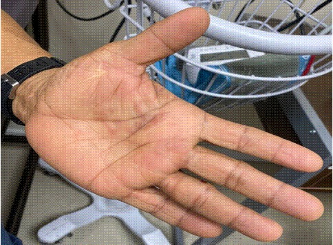
Figure 1. The initial presentation demonstrated an erythematous, serpiginous, slightly raised linear tract with irregular borders on the palmar surface, consistent with an actively migrating larval track. A smaller erythematous lesion with similar morphology is visible beneath the middle finger.
The patient initially sought treatment at an urgent care clinic, where he was diagnosed with presumed scabies and treated with permethrin 5% topical cream and oral prednisone. After 1 day without symptomatic improvement, he presented to our clinic for further evaluation. The localized serpiginous tract and recent sand exposure prompted reconsideration of the initial diagnosis.
Differential diagnosis. The differential diagnosis for a pruritic serpiginous eruption includes several infectious, inflammatory, and parasitic conditions. Larva currens typically migrates rapidly, advancing several centimeters per hour, and most commonly affects the perianal region or trunk. Tinea corporis was also considered, but the absence of annular scaling and central clearing made this diagnosis unlikely. Scabies commonly presents with burrows in characteristic locations, such as the interdigital spaces and flexural surfaces, and is often associated with intense nocturnal pruritus and involvement of multiple body sites. Other considerations included allergic contact dermatitis, irritant dermatitis, bacterial cellulitis, and cutaneous myiasis.
Final diagnosis. In this case, the combination of exposure history, lesion morphology, progressive serpiginous tract formation, and response to antiparasitic therapy strongly supported the diagnosis of cutaneous larva migrans (CLM).
Treatment and management. Given the characteristic serpiginous morphology of the lesion and recent sand exposure, cutaneous larva migrans was suspected. The patient was treated with a single oral dose of ivermectin (12 mg), calculated based on the recommended weight-based dosage of approximately 200 µg/kg (0.2 mg/kg) and topical triamcinolone acetonide 0.1% ointment for symptomatic relief of pruritus. Ivermectin was selected to optimize treatment adherence because of its simplified dosing regimen compared with the multi-day albendazole regimen. Within several days of initiating treatment, the patient reported significant improvement in pruritus, with only a few residual papules on the palmar surface of the hand (Figure 2). Two weeks later, a physical examination demonstrated flattening of the lesion with purpuric discoloration and mild desquamation.
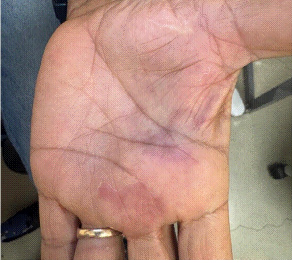
Figure 2. Two-week follow-up image of the left hand indicates flattening of the serpiginous lesion with purpuric discoloration and mild desquamation of the palmar skin after treatment.
Laboratory evaluation revealed mild peripheral eosinophilia, a nonspecific finding that may be seen in some parasitic infection but is not required for the clinical diagnosis of CLM. (Table 1). The full sequence of exposure, symptom progression, treatment, and clinical resolution took approximately 1 month. (Table 2).
Table 1. Complete blood count demonstrating mild peripheral eosinophilia. Test outside of normal range in bold.
|
Test |
Value |
Normal Range |
|
WBC (×10³/µL) |
8.1 |
4.0–10.0 |
|
RBC (×10⁶/µL) |
5.44 |
4.5–5.9 |
|
Hemoglobin (g/dL) |
14.1 |
13.5–17.5 |
|
Hematocrit (%) |
42.8 |
41–53 |
|
Platelets (×10³/µL) |
221 |
150–450 |
|
Absolute neutrophils |
3686 |
1500–8000 |
|
Absolute lymphocytes |
3094 |
1000–4000 |
|
Absolute eosinophils |
648 |
15–500 |
|
Absolute basophils |
41 |
0–100 |
Table 2. Clinical timeline of illness progression and treatment.
|
Time |
Clinical Event |
|
Day 0 |
Exposure to sand while performing yard work |
|
Day 1 |
Development of pruritic serpiginous rash on left palm |
|
Day 3 |
Urgent care visit; diagnosed as scabies |
|
Day 4 |
Clinic evaluation; CLM suspected |
|
Day 4 |
Oral ivermectin administered |
|
Day 7 |
Significant improvement in pruritus |
|
Week 2 |
Flattening of lesion with purpuric discoloration |
|
Month 1 |
Complete resolution |
Follow-up. At the patient’s 1-month follow-up, the patient reported complete resolution of symptoms with no residual rash or pruritus (Figure 3). The diagnosis of CLM was made based on exposure history, characteristic lesion morphology, evolution, and response to empiric antiparasitic therapy.
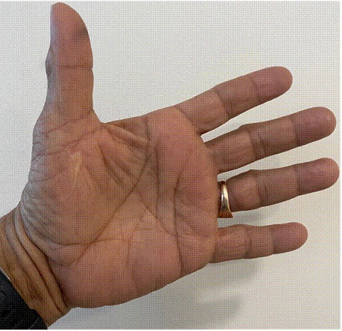
Figure 3. The palmar surface of the left hand shows complete resolution of the lesion with normal skin appearance and no residual erythema, papules, or purpura.
Discussion. Cutaneous larva migrans is a parasitic dermatosis caused by the migration of hookworm larvae, most commonly Ancylostoma braziliense or Ancylostoma caninum, within the epidermis. Humans are accidental hosts, and infection occurs when larvae penetrate the skin after contact with contaminated soil or sand. Although CLM is classically associated with tropical and subtropical travel, an increasing number of cases in the United States appear to be locally acquired, particularly in warm coastal regions, such as the southeastern United States.1 Environmental conditions, such as warm temperatures and sandy soil, facilitate larval survival, allowing transmission to occur even without international travel. The typical presentation consists of an intensely pruritic serpiginous track that migrates slowly across the skin, often advancing several millimeters to centimeters per day. Lesions most commonly occur on the feet, buttocks, or lower extremities due to direct ground contact. 2
Palmar involvement is less common and may delay diagnosis since clinicians often attribute pruritic eruptions of the hand to more common conditions, such as scabies, eczema, or contact dermatitis. In this case, the patient was initially misdiagnosed with scabies and treated with permethrin without improvement.
Although not universally present, peripheral eosinophilia may occur in CLM and can support the diagnosis when combined with clinical findings.3 Treatment options for CLM include oral ivermectin or albendazole, both of which are considered first-line therapy. Ivermectin offers the advantage of simplified dosing, typically administered as a single oral dose, whereas albendazole requires multi-day therapy.4 A comparative cohort study demonstrated cure rates of 90-100% with ivermectin compared with 77- 84.6% with albendazole, depending on treatment duration.2 Treatment with a single-dose oral ivermectin resulted in rapid symptom resolution and regression of the cutaneous lesion.5 Recent reports have raised concern that autochthonous CLM may occur outside traditional travel-associated contexts; proposed contributing factors include climate and environmental changes, though this case cannot establish those association.3,6,7
This case represents the importance of recognizing CLM even in patients without international travel and involving less common anatomical locations, such as the palm, where diagnosis may be delayed.
Conclusion. Our case highlights the importance of considering CLM in all patients presenting with pruritic, serpiginous eruptions, even in the absence of a history of international travel. Atypical lesion locations, such as the palm, and absence of travel history may contribute to diagnostic delay and misclassification as more common dermatologic conditions, including scabies or dermatitis. Careful attention to exposure history, lesion morphology, and migration pattern is essential for accurate diagnosis. Early recognition allows prompt treatment with antiparasitic therapy, which leads to rapid symptom resolution and prevents prolonged patient discomfort. As climate and environmental conditions continue to evolve, clinicians should remain aware that autochthonous CLM cases may increasingly occur in temperate regions of the United States.
Clinical Learning Points
- Cutaneous larva migrans can occur in the United States without international travel, particularly in the Southeast, where contaminated sand or soil may harbor hookworm larvae.
- Palmar involvement is uncommon and may lead to diagnostic delay, as lesions on the hand are frequently mistaken for scabies, contact dermatitis, or fungal infection.
- A serpiginous, slowly migrating erythematous tract with intense pruritus should prompt consideration of CLM, even when the anatomic location is atypical.
- Exposure history is critical, including yard work, beach activity, or contact with sand or soil potentially contaminated by animal feces.
- Single-dose oral ivermectin is an effective option for CLM and may provide rapid symptom relief and lesion regression.
AUTHORS:
Kristin Olenchak, MS-IV1 • Sophia Echevarria, MD2,3 • Sophia Kershner, MS-IV1 • Zafar Qureshi, MD3 • Colin A. Wilkinson, MD, PhD4 • Syed A. A. Rizvi, MD, PhD, MPH, MBA4,5
AFFILIATIONS:
1Nova Southeastern University Kiran C. Patel College of Osteopathic Medicine
2Universidad Mayor de San Simón, Cochabamba, Bolivia
3UMC Free Clinic, Miami Gardens, Florida, USA
4American International School of Medicine, Georgetown, Guyana, South America
5College of Biomedical Sciences, Larkin University, Miami, Florida, USA
CITATION:
Olenchak K, Echevarria S, Kershner S, Qureshi Z, Wilkinson CA, Rizvi SAA. A pruritic serpiginous palmar eruption in a Florida resident without international travel. Consultant. Published online June 29, 2026. DOI: 10.25270/con.2026.06.000001
DISCLOSURES:
The authors report no relevant financial relationships.
CONSENT FOR PUBLICATION:
Verbal consent has been obtained from the patient to publish his data.
ACKNOWLEDGEMENTS:
None.
CORRESPONDENCE:
Syed A. A. Rizvi, MD, PhD, MPH, MBA. Larkin University, 18301 N Miami Ave, Miami, FL 33169, USA (srizvi@larkin.edu)
References
- Crecelius E, Hickey P, Helfrich A. Cutaneous larva migrans within the United States Military Health System and association with International Travel. Open Forum Infectious Diseases. 2025;12(Supplement_1). doi:10.1093/ofid/ofae631.356
- Ryguła A, Kowalski M, Hryncewicz-Gwóźdź A, Maj J, Jankowska-Konsur A. Cutaneous larva migrans – case report and literature review. Family Medicine & Primary Care Review. 2023;25(3):367-370. doi:10.5114/fmpcr.2023.130099
- Can I, Yurekli A. Effect of global warming on dermatology practice: The increase in cases of cutaneous larva migrans in the eastern Black Sea region of Turkey. Journal of Cosmetic Dermatology. 2022;21(9):3929-3933. doi:10.1111/jocd.15128
- Johanis M, Cheema KS, Young PA, et al. Cutaneous larva migrans in the northeastern US. Dermatology Online Journal. 2023;29(4). doi:10.5070/d329461906
- Maxfield L, Crane JS. Cutaneous Larva Migrans. 2023 Jun 28. In: StatPearls [Internet]. Treasure Island (FL): StatPearls Publishing; 2026 Jan–. PMID: 29939528.
- Palaniappan V, Gopinath H, Karthikeyan K. Cutaneous larva migrans. Clinical and Experimental Dermatology. Published online August 7, 2025. doi:10.1093/ced/llaf375
- Liptáková M, Schreiberová A, Cellengová Z, Kožárová V, Štrkolcová G. The canine hookworm ancylostoma caninum: First confirmed evidence in a Dog in Central Europe: Epidemiological relevance or coincidence? Pathogens. 2025;14(12):1241. doi:10.3390/pathogens14121241
©2026 HMP Global. All Rights Reserved. Any views and opinions expressed are those of the author(s) and/or participants and do not necessarily reflect the views, policy, or position of Consultant360 or HMP Global, their employees, and affiliates.