


A Mimic of Neonatal Mastitis: Superficial Cellulitis in Physiologic Neonatal Gynecomastia
Introduction. A 2-week-old afebrile male infant presented to the emergency department with a 2-day history of unilateral breast erythema and subareolar swelling.
History. The patient’s father initially noted mild enlargement of the right breast, which progressed over 48 hours with increasing size and erythema, prompting presentation to the emergency department. The patient was feeding well on breast milk and formula, with normal urine output, bowel movements, sleep, activity level, and appetite. The patient remained afebrile, with no vomiting, diarrhea, respiratory distress, or sick contacts reported. The parents did not attempt any interventions at home.
The infant was born at 39 weeks and 4 days’ gestation via uncomplicated spontaneous vaginal delivery to a 32-year-old G2P2 mother. Maternal prenatal laboratory studies were unremarkable, including negative screening for group B Streptococcus, hepatitis B, HIV, and syphilis. There were no perinatal complications, and the neonatal course was uncomplicated.
On presentation, vital signs were within normal limits for age. The infant’s rectal temperature was 36.4°C, his heart rate was 141 beats per minute, his respiratory rate was 56 breaths per minute, oxygen saturation was 97% on room air, and he weighed 3.3 kg. The patient was initially sleeping but readily arousable, quickly becoming alert and appropriately responsive during examination.
Physical examination. The physical examination revealed a well-appearing neonate with no signs of systemic illness. The anterior fontanelle was open and soft. The cardiopulmonary and abdominal examinations were normal. The genital examination demonstrated normal male genitalia with bilaterally descended testes and mild bilateral hydroceles.
The examination of the chest revealed a firm, mobile subareolar mass beneath the right areola with overlying erythema, warmth, and tenderness to palpation (Figure 1A-C). We also palpated a smaller, non-tender subareolar mass beneath the left areola without associated erythema or warmth. Following gentle palpation, a small amount of nipple discharge was expressed from the right nipple. The remainder of the chest and dermatologic examination were unremarkable. Given our concern for neonatal mastitis, the patient was admitted to the pediatric service for observation, further evaluation, and initiation of empiric antimicrobial therapy.
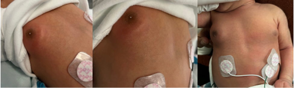
Figure 1A-C. Erythema overlying right subareolar gynecomastia, with minimal white nipple discharge (A, B). Bilateral subareolar gynecomastia, right greater than left (C).
Breast ultrasonography demonstrated a circumscribed, heterogeneous, hypoechoic subareolar structure measuring 29 × 9 × 24 mm in the right breast, with mild internal vascularity. A similar but smaller structure was identified in the left breast, measuring 20 × 5 × 20 mm. No fluid collection, abscess formation, or deep parenchymal infection was identified (Figure 2A-B).
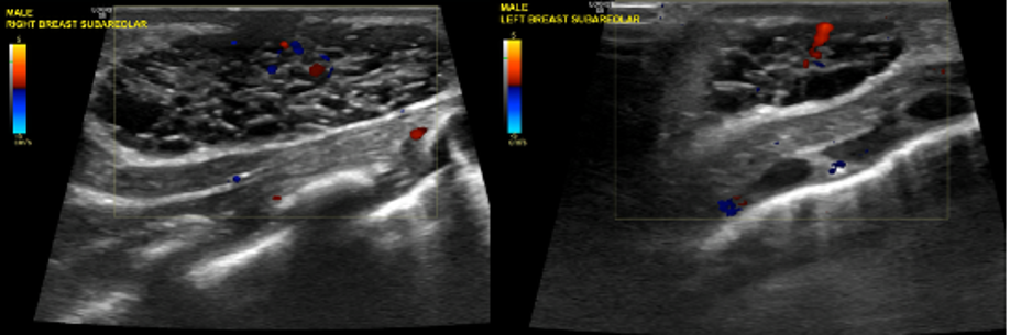
Figure 2A-B. Ultrasound images of the right (A) and left (B) breasts show circumscribed heterogeneous hypoechoic subareolar structures, right greater than left, with mild internal vascularity, consistent with physiologic neonatal gynecomastia and without evidence of abscess formation.
These findings were consistent with physiologic neonatal bilateral gynecomastia, with superimposed superficial soft-tissue inflammation of the right breast.
Diagnosis. Our team, led by the pediatric attending on call, then made the diagnosis of physiologic neonatal gynecomastia with superimposed right-sided superficial cellulitis.
Differential diagnoses. Several diagnoses were considered based on this infant's clinical presentation. Neonatal mastitis was initially suspected due to unilateral breast erythema, warmth, tenderness, and progressive swelling.1 However, the absence of fever or systemic symptoms, normal inflammatory markers, stable vital signs, and reassuring ultrasonographic findings made a deep breast infection less likely.2
Secondary differential diagnosis included galactoceles versus breast abscess. Galactoceles are benign cystic breast lesions containing milk or milk-like fluid and are most often encountered during or shortly after lactation.3 Breast abscess formation is a known complication of neonatal mastitis.4 Ultrasonography demonstrated no fluid collection or cystic lesion, making an abscess or galactocele less likely.5
The absence of systemic illness, evidence of no deep breast involvement, and localized erythema and tenderness confined to the skin and subcutaneous tissues ultimately supported a diagnosis of superficial cellulitis.
Treatment and management. Upon admission, we counseled the parents to avoid squeezing, massaging, or manipulating either breast, as these actions may exacerbate inflammation or introduce bacteria by disrupting the skin barrier.4 We initiated supportive care and close clinical observation, which continued throughout the entire hospitalization.
We initiated intravenous clindamycin at approximately 7.5 mg/kg per dose (25 mg every 8 hours) because of concern for neonatal mastitis. The patient received the first dose on the evening of admission/hospital day 1. We gave him the second dose on the morning of hospital day 2.
Midday on hospital day 2, ultrasonography demonstrated no abscess, deep infection, or fluid collection, and laboratory results demonstrated no elevations in inflammatory markers or leukocytes. As the patient was clinically stable with reassuring vital signs, the antimicrobial therapy was de-escalated to oral cephalexin, dosed at 25 mg/kg per dose, administered as 80.875 mg 3 times daily, with a planned 9-day course.1
Outcome and follow-up. The infant remained afebrile and hemodynamically stable with normal feeding, urine output, and stooling throughout the entire hospital course. The blood cultures showed no growth at 24 or 48 hours. Repeat laboratory values on hospital day 2 remained within normal limits.
On hospital day 2, the patient’s physical examination was notable for a mild decrease in right subareolar swelling and significant decreases in erythema, warmth, and tenderness compared with the initial presentation. We did not note any changes in the left breast or in the left subareolar swelling. By hospital day 3, there was full resolution of right breast erythema, warmth, and tenderness. The subareolar swelling was still present, right greater than left, unchanged since the previous examination.
The patient was discharged on hospital day 3 in stable condition with instructions to complete the 9-day course of oral cephalexin 80.875 mg 3 times daily, and to follow up with his outpatient pediatrician within 3-5 days. The parents were advised to return for medical evaluation if fever, worsening swelling, purulent discharge, or systemic symptoms developed. The patient did not present to the outpatient follow-up appointment. Since his discharge, there have been no documented presentations or readmissions for this complaint.
Discussion. Physiologic neonatal gynecomastia is a benign and self-limited condition resulting from transplacental passage of maternal estrogens and subsequent neonatal pituitary stimulation.6,7 It occurs in up to 60% to 90% of newborns and may present with bilateral or unilateral subareolar breast enlargement, occasionally accompanied by milky nipple discharge or neonatal galactorrhea.8 The condition typically resolves spontaneously over weeks to months without intervention.9
Neonatal mastitis, in contrast, is an uncommon but potentially serious infection that most often presents within the first 2 to 4 weeks of life.4 It is characterized by unilateral breast swelling, erythema, warmth, tenderness, and occasional fever or systemic signs of illness.1,2 Staphylococcus aureus is the most commonly implicated bacterial pathogen.2,3 Due to the risk of bacteremia and further progression to abscess formation, neonatal mastitis has traditionally been treated with parenteral antibiotics, with the addition of surgical drainage when indicated by the presence of abscess formation on ultrasonography.3,4
This case illustrates the diagnostic challenge that arises when physiologic neonatal gynecomastia is accompanied by localized inflammatory changes. This patient demonstrated clinical features concerning mastitis, including erythema, tenderness, and progressive swelling. However, the diagnosis of superficial cellulitis rather than neonatal mastitis was supported by the overall clinical presentation, normal inflammatory markers, and ultrasonographic findings demonstrating no evidence of breast parenchymal infection or abscess.9,10
An additional teaching point highlighted by this case is the importance of avoiding manual expression or manipulation of hypertrophied neonatal breast tissue. Mechanical manipulation may disrupt the skin barrier and introduce bacteria, increasing the risk of secondary infection.4 Parental education regarding avoidance of breast manipulation is, therefore, a critical component of management.
Ultrasonography played a pivotal role not only in establishing the correct diagnosis but also in guiding appropriate treatment. The absence of fluid collection or cystic structures indicated that no true parenchymal infection was present, making neonatal mastitis less likely.5 The presence of bilateral, well-circumscribed, heterogeneous hypoechoic subareolar structures on ultrasound supported a diagnosis of physiologic neonatal gynecomastia. These ultrasonography findings, combined with this patient's clinical presentation, indicated a superficial soft-tissue infection overlying benign neonatal breast tissue.
Most importantly, the utilization of ultrasonography during the initial diagnostic evaluation can facilitate early and accurate diagnostic clarification, distinguishing benign physiologic breast hypertrophy with overlying superficial cellulitis from neonatal mastitis. A key consideration between these similarly presenting conditions is also the substantial difference in the quantity and duration of antibiotic therapy required. In this case, following inpatient observation and clinical improvement, antimicrobial therapy was de-escalated to oral antibiotics for the remainder of treatment. Timely and accurate diagnosis can prevent prolonged observation and reduce overtreatment to minimize the risks and potential adverse effects associated with antimicrobial therapy in neonates.2
Although superficial cellulitis associated with neonatal gynecomastia is infrequently described in the literature, it remains an important diagnostic consideration. Within this case, ultrasonography excluded a drainable abscess, which in combination with a reassuring clinical exam and normal inflammatory markers, supported the diagnosis of superficial cellulitis over neonatal mastitis.
Conclusion. This case demonstrates a stepwise approach to neonatal breast pathology that balances patient safety with antibiotic stewardship and emphasizes the importance of ultrasonography when distinguishing physiologic neonatal gynecomastia from neonatal mastitis. Although the patient demonstrated clinical improvement during hospitalization and was discharged in stable condition, long-term clinical resolution was not confirmed.
AUTHORS:
Alissa Reed, BS1 • Erin Kim, BS2 • Scott Hushaw, BS2 • Hila Yousefi, BS1 • Thiagarajan Nandhagopal, MD3
AFFILIATIONS:
1College of Osteopathic Medicine of the Pacific-Northwest, Western University of Health Sciences, Lebanon, OR, USA
2College of Osteopathic Medicine of the Pacific, Western University of Health Sciences, Pomona, CA, USA
3Kern Medical Center, Bakersfield, CA, USA
CITATION:
Reed A, Kim E, Hushaw S, Yousefi H, Nandhagopal T. A mimic of neonatal mastitis: superficial cellulitis in physiologic neonatal gynecomastia. Consultant. 2026;66(8). doi: 10.25270/con.2026.08.000001
DISCLOSURES:
The authors report no relevant financial relationships.
CONSENT FOR PUBLICATION:
Consent has been obtained from the patient’s parents to publish his data.
ACKNOWLEDGEMENTS:
None.
CORRESPONDENCE:
Alissa Reed, BS. College of Osteopathic Medicine of the Pacific-Northwest, Western University of Health Sciences. 200 Mullins Dr, Lebanon, OR 97355 (email: Alissa.reed@westernu.edu)
References
- Kaplan RL. Neonatal mastitis: clinical presentation and approach to evaluation and management. Pediatr Emerg Care. 2024;40(12):920-921. doi:10.1097/PEC.0000000000003281
- Kaplan RL, Cruz AT, Michelson KA, et al. Neonatal mastitis and concurrent serious bacterial infection. Pediatrics. 2021;148(1). doi:10.1542/peds.2021-051322
- de Chadarévian JP, Arthur LG, Rezvani GA, Duke DS, Davis WJ, Faerber EN. The galactocele of male infants: an intriguing entity. Study and reflection about a case, with review of the literature. Pediatr Dev Pathol. 2011;14(2):144-148. doi:10.2350/10-05-0827-CR.1
- Stricker T, Navratil F, Sennhauser FH. Mastitis in early infancy. Acta Paediatr. 2005;94(2):166-169. doi:10.1080/08035250410022576
- Borders H, Mychaliska G, Gebarski KS. Sonographic features of neonatal mastitis and breast abscess. Pediatr Radiol. 2009;39(9):955-958. doi:10.1007/s00247-009-1310-x
- Sansone A, Romanelli F, Sansone M, Lenzi A, Di Luigi L. Gynecomastia and hormones. Endocrine. 2017;55(1):37-44. doi:10.1007/s12020-016-0975-9
- Hiba J, Pozo ED, Genazzani A, et al. Hormonal mechanism of milk secretion in the newborn. J Clin Endocrinol Metab. 1977;44(5):973-976. doi:10.1210/jcem-44-5-973
- Dickson G. Gynecomastia. Am Fam Physician. 2012;85(7):716-721.
- Cakan N, Kamat D. Gynecomastia: evaluation and treatment recommendations for primary care providers. Clin Pediatr (Phila). 2007;46(6):487-490. doi:10.1177/0009922806294800
- Stocker M, van Herk W, El Helou S, et al. C-reactive protein, procalcitonin, and white blood count to rule out neonatal early-onset sepsis within 36 hours: a secondary analysis of the Neonatal Procalcitonin Intervention Study. Clin Infect Dis. 2021;73(2). doi:10.1093/cid/ciaa876
- Norman-Bruce H, Umana E, Mills C, et al. Diagnostic test accuracy of procalcitonin and C-reactive protein for predicting invasive and serious bacterial infections in young febrile infants: a systematic review and meta-analysis. Lancet Child Adolesc Health. 2024;8(5):358-368. doi:10.1016/S2352-4642(24)00021-X