
Authors:
Tiffany Leung, MD
Department of Pediatrics, University of California, Irvine, School of Medicine, Irvine, California and Children’s Hospital of Orange County, Orange, California
Sunil Kamath, MD
Department of Pediatric Pulmonology, Children’s Hospital of Orange County, Orange, California
Geetha Puthenveetil, MD
Department of Pediatric Hematology/Oncology, Children’s Hospital of Orange County, Orange, California
Citation:
Leung T, Kamath S, Puthenveetil G. Infantile pulmonary hemangioma [published online January 22, 2018]. Consultant for Pediatricians.
A 2-month-old boy was admitted to the hospital for a 2-day history of respiratory distress.
Diagnostic imaging. A chest radiograph showed near opacification of the right lung with a slight leftward mediastinal shift. Contrast enhanced computed tomography (CT) of the chest showed a large vascular mass in the right hemithorax and upper abdomen (Figure 1). Magnetic resonance imaging (MRI) of the chest and abdomen revealed 2 left upper quadrant abdominal masses. The pulmonary mass was biopsied and results eventually confirmed it to be an infantile hemangioma.
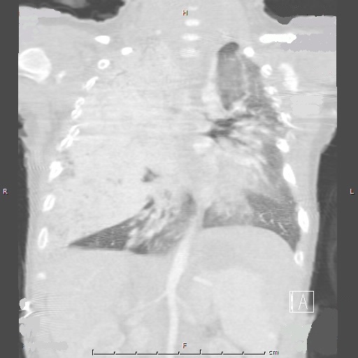
Figure 1: Computed tomography of the chest with contrast performed at 2 months of age shows enhancing masses in the right lung and upper abdomen.
Treatment. He was treated with interferon alfa and prednisolone for 4 years.
Follow-up. A CT scan of the chest was repeated approximately 1 year later and showed more cystic appearing lesions and smaller intrathoracic and abdominal masses compared to previous scans. MRI scans of the chest obtained annually during therapy demonstrated a gradual decrease in size of the hemangiomas. A follow-up CT scan of the chest performed at age 7 showed extensive centrilobular emphysema and dysplasia of most of the right lung (Figure 2). Despite the significant amount of dysplastic lung tissue, the patient did not have any recurrent illnesses or medication needs, and he remains well from a pulmonary standpoint.
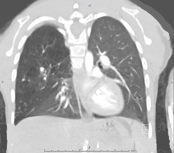
Figure 2: Computed tomography of the chest with contrast performed at 7 years of age shows extensive centrilobular emphysema of the majority of the right lung.
Discussion. Infantile hemangioma is a common benign vascular tumor with characteristic phases of growth, proliferation, and involution. The prevalence rate is approximately 10% of all infants.1 Most lesions are cutaneous and affect the head, neck, or trunk.2 Pulmonary involvement is extremely rare with only a few reported cases to date.3-5 Interestingly, this patient had also presented a few years before propranolol had become the widely accepted treatment for infantile hemangioma, hence he was treated with corticosteroids and interferon alfa. To our knowledge, cases of lung dysplasia with emphysematous changes subsequent to corticosteroid and interferon alfa administration for pulmonary hemangioma have never before been reported.
In patients with pulmonary hemangioma, it is important to emphasize the importance of long term follow-up with pulmonology and hematology specialists. Repeated imaging must be obtained during the course of therapy to demonstrate response to treatment. Other diagnostic tests, including polysomnography and bronchoscopy, can be helpful. Although infantile hemangiomas are benign, post-treatment complications are bound to occur especially among patients on long term therapy. Involution of the lesion is likely to leave behind significant scarring and may alter the architecture of the lung, as in the case of our patient. Obstructive or restrictive lung disease may develop so routine pulmonary function testing is also recommended.
In conclusion, hemangioma should be considered as an etiology for respiratory distress in an infant and especially if a pulmonary mass is visualized on imaging results. Treatment options have evolved in the past decade and now medications with less severe side effects are used. However, providers still need to closely monitor the clinical course with routine imaging and diagnostic studies to ensure resolution of the lesions.