
Worsening Swelling and Pain on the Side of a Girl’s Face
A mother brought her otherwise healthy 22-month-old daughter to a pediatric clinic with concern about swelling and pain over the left side of the girl’s face that had been progressively worsening over the last 2 days. The mother had noticed a small bump on the left side of the child’s face 2 days ago, and she initially thought it was secondary to trauma that the girl had sustained while playing with her siblings.
The child had no fever or symptoms of a cold, and her appetite and activity level were normal. She had no history of recent illnesses, insect bites or stings, or bruising in the area. She had not fallen recently, and she had no history of travel. The rest of the girl’s history was noncontributory.
Physical examination findings, including vital signs, were normal, except that the left side of her face was swollen, warm to the touch, and tender, but with no induration or fluctuance. She also had a palpable, 1-cm, left anterior cervical lymph node, which was nontender, mobile, and without increased warmth of the overlying skin.

What might be causing these symptoms?
(Answer and discussion on next page)
Answer: Infection of the ear pit
The girl received a diagnosis of an infection of the ear pit.
Incidence and Epidemiology
Ear pits are known by many different names, including preauricular sinuses, congenital preauricular fistulas, and congenital auricular fistulas. They are caused by congenital malformations of the first and second branchial arches and most often are inherited in an incomplete autosomal dominant pattern, although they also can arise spontaneously.1 These sinuses typically have small external orifices on the surface of the skin, but the course of the duct can extend into the vicinity of the facial nerve and can even continue into the parotid gland.2
In the United States, the incidence of ear pits is estimated to be approximately 0.9%; internationally, ear pits appear to be most prevalent in areas of Asia and Africa, where they are found in from 4% to 10% of the population.1 Consequently, ear pits are more prevalent in African Americans and Asian Americans than in white Americans.
Ear pits are bilateral in 25% to 50% of cases, and unilateral cases most often are found on the left ear.1 Males and females appear to be equally affected. Preauricular sinuses are associated with a number of conditions, including third molar impactions, renal malformations/renal agenesis, cleft palate, Treacher Collins syndrome, hemifacial microsomia, and branchio-oto-renal syndrome.3
Presentation
Ear pits most frequently present at birth but also can become visible later in life. Ear pits most often are seen on physical examination as a nodule, dent, or dimple located at the anterior margin of the ascending limb of the helix, but they can be found anywhere on the surface of the external ear.1
In most patients with them, ear pits are asymptomatic, but some patients can present with chronic transient purulent drainage from the orifice. Ear pits with chronic drainage are predisposed to frequent infections and may progress to facial cellulitis or ulcerations. Often, the cellulitis or ulcer is treated medically, and the underlying anatomic defect of the ear pit is never appreciated, consequently setting up the patient for recurrent infections. Scarring and disfigurement can result from an infection of an ear pit. 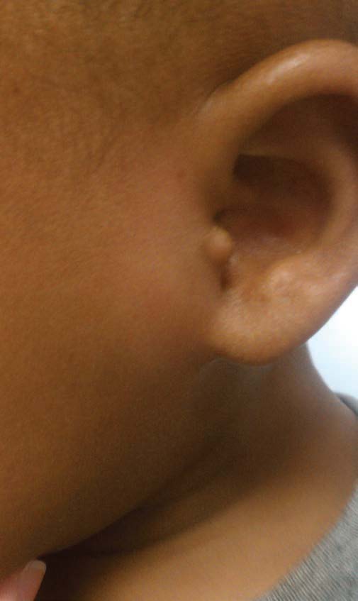
Management
People with preauricular pits have an increased risk of permanent hearing loss compared with the general population. A retrospective study of 68,484 infants over 7.5 years found a 0.93% incidence of preauricular skin tags and/or pits.4 Infants with preauricular skin tags or ear pits had an 8 in 1,000 prevalence of permanent hearing impairment compared with 1.5 in 1,000 in infants without tags or pits.
Although there is an association between preauricular pits and renal anomalies, the incidence of renal anomalies in those with an isolated preauricular pit is comparable with that of the general population, and renal ultrasonography is not indicated in the routine evaluation of such an infant.5 Renal ultrasonography is recommended in patients with a preauricular pit or tag who also have another dysmorphic feature, a family history of deafness or auricular or renal malformations, and/or a maternal history of gestational diabetes.5
Surgery is indicated only in cases of recurrent infection of the ear pit or squamous discharge from it.1 Surgery should include excision of the pit, the squamous-lined cyst, and the cartilage at the root of the helix as a whole to prevent recurrence.6
Sharda Udassi, MD, is an assistant professor in Division of Pediatric Hospital Medicine at Shands Children’s Hospital at the University of Florida College of Medicine in Gainesville.
Nancy George, MD, is a second-year pediatrics resident at the University of Florida College of Medicine in Gainesville.
Zachary Boucher is a medical student at the University of Florida College of Medicine in Gainesville.
Sanjeev Y. Tuli, MD, is a professor of pediatrics, associate chair for Clinical Affairs and Community Relations, and Chief of the Division of General Academic Pediatrics at the University of Florida College of Medicine in Gainesville.
References
1. Scheinfeld NS, Silverberg NB, Weinberg JM, Nozad V. The preauricular sinus: a review of its clinical presentation, treatment, and associations. Pediatr Dermatol. 2004;21(3):191-196.
2. Leung AKC, Robson WLM. Association of preauricular sinuses and renal anomalies. Urology. 1992;40(3):259-261.
3. Tan T, Constantinides H, Mitchell TE. The preauricular sinus: a review of its aetiology, clinical presentation and management. Int J Pediatr Otorhinolaryngol. 2005;69(11):1469-1474.
4. Roth DA, Hildesheimer M, Bardenstein S, et al. Preauricular skin tags and ear pits are associated with permanent hearing impairment in newborns. Pediatrics. 2008;122(4):e884-e890.
5. Kugelman A, Tubi A, Bader D, Chemo M, Dabbah H. Pre-auricular tags and pits in the newborn: the role of renal ultrasonography. J Pediatr. 2002;141(3):388-391.
6. Prasad S, Grundfast K, Milmoe G. Management of congenital preauricular pit and sinus tract in children. Laryngoscope. 1990;100(3):320-321.