
Skin Disorders in Older Adults: Benign Cellular Proliferations
ABSTRACT: Among the benign cellular proliferations that can occur in the elderly are fibrous papules, scars, skin tags, sebaceous hyperplasia, and lichen planus–like keratosis. Some of these growths can be confused with malignant conditions. For example, fibrous papules can resemble basal cell carcinomas. If any confusion exists, perform a shave biopsy and submit the specimen for histopathological examination. A pitfall in the diagnosis of scars is the failure to recognize mimics, particularly metastatic cancers that manifest as linear plaques. Lichen planus–like keratosis is a solitary keratotic papule or plaque that clinically resembles an actinic keratosis, Bowen disease, or basal cell carcinoma.
A variety of cellular proliferations resulting from cell damage or hormonal, cytokine, and inflammatory mediators can occur in the skin. Fibroblastic proliferations, such as scars, keloids and fibrous papules, do not resemble normal skin, but they are not actually pathologic. Other benign growths that can occur in the elderly are skin tags, sebaceous hyperplasia, and lichen planus–like keratosis. In this article, I will describe and illustrate these processes.
 FIBROUS PAPULES
Fibrous papules are benign and most commonly arise on the nose. The cause is unknown. They usually manifest as firm papules on the nasal alae (Figure 1). In most cases, they are solitary, but sometimes more than one papule is present. Occasionally, fibrous papules appear on the cheeks, chin, neck, and, rarely, lip or forehead. The papules are usually dome-shaped, firm, flesh to pink to red in color, shiny, and 1 to 6 mm in diameter. Rarely, fibrous papules are sessile, polypoid, verrucous, or papillomatous.
Histologically, they demonstrate dermal stellate cells that contain factor XIIIa and vimentin, a fibrotic stroma, and dilated blood vessels. Occasionally, a sparse inflammatory cell infiltrate of lymphocytes is present.1
Fibrous papules can be confused with basal cell carcinomas and intradermal nevi. If any confusion exists, perform a shave biopsy and submit the specimen for histopathological examination. Fibrous papules tend not to reoccur after shave biopsy removal.
 SCARS, HYPERTROPHIC SCARS, AND KELOIDS
The elderly undergo a variety of operations, including open heart surgery, hip surgery, and various types of gastrointestinal surgery. The surgical scars that result from these procedures are less noticeable and extensive in older adults than they are in younger persons. Usually, surgical scars in the elderly, particularly in areas without skin tension, are soft and slightly lighter in hue than the surrounding skin (Figure 2).2 Few of these surgical scars, even in persons of color, become raised or fibrotic within the site of the scar (hypertrophic scars) (Figure 3) or extend beyond the scar margins (keloids) (Figure 4).
Keloids are lesions that occur in response to injury and extend beyond the margins of a site of injury or surgery. They have little surface change, are firm or rubbery, and are more common in darker skinned persons. They can be pruritic or painful. They are most common in the young (those between the ages of 10 and 30) but can occur in persons of any age.3 I treat them with injections of 1 to 3 mg/cc of intralesional triamcinolone suspension to decrease the size and coincident pruritus of the lesions.
A pitfall in the diagnosis of scars is the failure to recognize the mimics of scars, particularly metastatic cancers. I have encountered linear plaques in the breasts of patients with a history of breast cancer that biopsy revealed to be recurrent breast cancer and not keloids. Any keloid in an older adult that seems atypical should be biopsied because keloids typically do not develop in the elderly. In addition, certain inflammatory diseases, such as psoriasis, can arise in sites of injury and along scars; this is known as the Koebner phenomenon.4 In the case of psoriasis, it is referred to as koebnerized psoriasis (Figure 5).
 SKIN TAGS
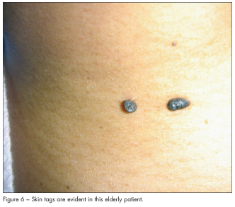
Skin tags are growths of tissue that are related to genetics, diabetes, and obesity. They tend to become more common with age. They appear as flesh-colored to brown papules that can be on stalks (Figure 6). They can be smooth or warty but are almost always soft to palpation. I remove them with gradle scissors after injecting lidocaine at their base.
 SEBACEOUS HYPERPLASIA
Sebaceous hyperplasia is a common, benign condition in the elderly that is characterized by an increase in sebaceous glands. It can be related to sun damage and rosacea. Sebaceous hyperplasia manifests as yellowish, soft, small papules on the face (particularly eyelids, nose, cheeks, and forehead) (Figure 7); it can occur as a single lesion or as multiple lesions. The condition does not need to be treated; however, sparing topical application of 20% to 35% trichloroacetic acid can clear it or make it less apparent.
 LICHEN PLANUS–LIKE KERATOSIS
Lichen planus–like keratosis (LPLK) is a solitary keratotic papule or plaque that clinically resembles an actinic keratosis, lentigo, Bowen disease, or a basal cell carcinoma. LPLK is also known as a benign lichenoid keratosis and involuting lichenoid plaque. LPLK typically occurs on sun-exposed areas of the arms, legs, and presternal region. It is usually purple and sometimes has a halo of erythema and a scaly surface (Figure 8).
As the name suggests, the histology of an LPLK resembles that of lichen planus, although there may be subtle differences.5 An LPLK probably represents a regressing lentigo or seborrheic keratosis and, if sought for, a remnant of these former proliferations may be found. A skin biopsy helps distinguish between this lesion and lichen planus. The most helpful clue is the clinical presentation. If multiple lesions are present, lichen planus is the probable diagnosis. I tend to use cryotherapy on LPLKs and biopsy them to rule out basal cell cancer or squamous cell cancer.
1. Bansal C, Stewart D, Li A, Cockerell CJ. Histologic variants of fibrous papule. J Cutan Pathol.2005;32(6):424-428.
2. Bayat A, McGrouther DA, Ferguson MWJ. Skin scarring. BMJ. 2003;326(7380):88-92.
3. Murray JC, Pollack SV, Pinnell SR. Keloids: a review. J Am Acad Dermatol. 1981;4(4):461-470.
4. Weiss G, Shemer A, Trau H. The Koebner phenomenon: review of the literature. J Eur Acad Dermatol Venereol. 2002;16:241-248.
5. Morgan MB, Stevens GL, Switlyk S. Benign lichenoid keratosis: a clinical and pathologic reappraisal of 1040 cases. Am J Dermatopathol. 2005;27(5):387-392.