
Peer Reviewed
Bullous Tinea
AUTHORS:
Anna Tappel, BS
University of Virginia School of Medicine, Charlottesville, Virginia
Barrett Zlotoff, MD
Department of Dermatology, University of Virginia Health System, Charlottesville, Virginia
CITATION:
Tappel A, Zlotoff B. Bullous tinea [published online October 12, 2018]. Consultant360.
An 8-year-old girl who was on maintenance chemotherapy for acute lymphoblastic leukemia presented for evaluation of a pruritic lesion on her left hand that had failed to respond to treatment with topical triamcinolone.
On physical examination, annular erythematous pustules were present on the ventral and dorsal aspects of her left hand (Figure). Her brother had an annular scaly rash on his scalp with acute occipital lymphadenopathy.
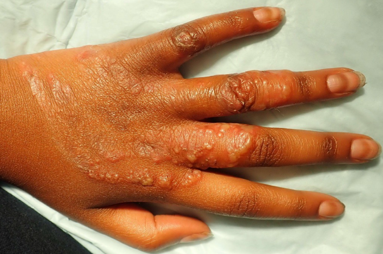
This is bullous tinea, an uncommon cutaneous dermatophyte infection that is most often caused by Trichophyton rubrum or Trichophyton mentagrophytes.1 Bulla formation is thought to be secondary to a delayed hypersensitivity provoked by the presence of dermatophyte antigens.2
In children, signs of dermatophyte infection can be nonspecific and are frequently misdiagnosed as psoriasis, atopic dermatitis, contact dermatitis, dyshidrotic eczema, herpetic infection, cellulitis, or impetigo.1,3 The strongest predisposing factor for bullous tinea is likely a history of tinea infection in a family member.4
The diagnosis should be confirmed with microscopic examination of a potassium hydroxide (KOH) preparation, and a fungal culture or biopsy should be considered if KOH test results are negative but clinical suspicion for bullous tinea remains high.
Both topical and systemic antifungal agents are therapeutic options, depending on the drug safety profile and the patient’s clinical disease involvement and disease duration.
Due to this patient’s immunosuppression and concerns for medication interactions, she was treated with 2% ketoconazole shampoo and 200 mg of fluconazole orally for 30 days, which resulted in clinical resolution of the rash at her 1-month follow-up appointment.
References:
- Romano C, Rubegni P, Ghilardi A, Fimiani M. A case of bullous tinea pedis with dermatophytid reaction caused by Trichophyton violaceum. Mycoses. 2006;49(3):249-250.
- Svejgaard E, Christiansen AH, Stahl D, Thomsen K. Clinical and immunological studies in chronic dermatophytosis caused by Trichophyton rubrum. Acta Derm Venereol. 1984;64(6):493-500.
- Sweeney SM, Wiss K, Mallory SB. Inflammatory tinea pedis/manuum masquerading as bacterial cellulitis. Arch Pediatr Adolesc Med. 2002;156(11):1149-1152.
- Neri I, Piraccini BM, Guareschi E, Patrizi A. Bullous tinea pedis in two children. Mycoses. 2004;47(11‐12):475-478.