
Peer Reviewed
Amoebic Liver Abscess: Case Report and Updates to Management
Introduction. A 56-year-old man presented to the emergency department of a large academic hospital in Dallas, TX with sudden onset abdominal pain the night prior to presentation.
History. The patient’s medical history was remarkable for deep venous thrombosis complicated by pulmonary embolism (PE) while undergoing orthopedic intervention in his right knee for tendon rupture 5 years prior to admission. The history of the present illness was pertinent for palpitations and chills for 1 week, but negative for fever, diarrhea, nausea, vomiting, unintentional weight loss, and loss of appetite.
The patient recently traveled to Mexico 1 month prior to symptom onset. The patient has worked as a cleaner in a company for portable public restrooms for more than 3 years. He reported wearing appropriate personal protective equipment during his work.
Upon arrival at the hospital, the patient had a temperature of 37.3 °C, pulse of 67 beats per minute, and blood pressure of 112/75 mmHg. The patient’s abdominal examination indicated mild discomfort from palpation, which was most significant in the right upper abdominal quadrant; the patient otherwise did not exhibit guarding nor was noted to have hepatosplenomegaly. Cardiovascular, respiratory, and neurological examinations were unremarkable. No jaundice was noted on examination of the skin and sclera.
Diagnostic Testing. Laboratory findings were significant for mild leukocytosis with a white blood cell count of around 11,000 cells/μL with neutrophilic dominance, and an eosinophilic count that was within normal limits. The patient’s hepatic enzymes were also within normal limits. An ultrasound of the gallbladder was negative for an acute biliary process. The ultrasound of the liver was remarkable for heterogenous right hepatic mass measuring up to 6.6 cm (about 2.6 in) with associated peripheral vascularity and no significant cystic component, which is most concerning for a neoplastic process. Computed Tomography (CT) of the abdomen and pelvis was performed for further characterization (Figure 1). The imaging confirmed the diagnosis of a hepatic abscess however, we did not know whether the etiology was amoebic or pyogenic.
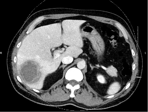
Fig. 1. Initial CT scan showing hepatic abscess.
Differential diagnoses. Initially, the patient was thought to have cholecystitis given the nature of his pain and subjective fever. After the ultrasound result was obtained, there was concern that he had a malignant tumor of the liver. However, CT confirmed that he had a hepatic abscess. A bacterial infection was higher on the differential, as amoebic infections are generally uncommon in the United States.
Treatment and management. The patient was admitted to the hospital ward for further diagnosis and management. Given the unclear etiology initially, antimicrobial treatment was held until cultures from the abscess were obtained. On the second day of admission, interventional radiology inserted a 10 Fr drain into the hepatic abscess and drained 10 cc of fluid. The drain was left in place for continued drainage, and fluid cultures were obtained. Empiric treatment was initiated with 2 grams once a day of ceftriaxone and 500 mg three times daily of metronidazole. On day 7 of hospitalization, stool Ova and Parasite studies were positive for Entamoeba histolytica and serum Entamoeba histolytica was also positive, confirming diagnosis of amoebic abscess. The patient was then started on metronidazole 750 mg three times day for a 10-day course, which was followed by luminal treatment with paromomycin at a dose of 750 mg three times daily for 10 days.
On day 11 of hospital admission, output from the hepatic drain was very minimal, and the drain was removed.
His hospital stay was further complicated by a PE that was incidentally noted on repeat CT scan of the abdomen. At that time, the patient had reported mild chest pain. The patient was hemodynamically stable at that time and denied change in symptoms. He was ambulatory throughout his hospital stay and was on an appropriate dose of enoxaparin sodium for deep venous thrombosis prophylaxis. The chest CT angiography confirmed the PE, and the patient was treated initially with heparin drip and transitioned to rivaroxaban 15 mg BID for 21 days followed by 20 mg QD for a total of 6 months due to history of recurrent thromboembolism at discharge on day 11.
On discharge from the hospital, the patient’s symptoms had significantly improved with the resolution of his abdominal pain. A follow-up CT of the abdomen was arranged, and he was scheduled for a hospital follow-up visit with his primary care doctor clinic 3 weeks later.
Outcome and follow-up. On follow-up in the clinic 3 weeks post-discharge, a repeat CT of the abdomen and pelvis noted an interval decrease in the size of the phlegmonous change in segment 6 of the liver with central liquefied abscess of less than 1 cm in diameter from a size of 5.8 x 6.1 x 6.0 cm in an interval of 4 weeks. His symptoms had resolved, and he had completed his course of antimicrobial treatment.

Fig. 2. CT scan at 4-week follow-up showing significant decrease in size of the abscess.
Discussion. Amoebiasis, a gastrointestinal protozoan disease caused by Entamoeba histolytica, is the second most common cause of death from parasitic disease in the world behind only malaria.1 Amoebic liver abscess (ALA) is the most common extraintestinal manifestation of this disease and has been observed in 3% to 9% of cases.2 There are no formal guidelines for managing ALA, but generally, treatment and management involve metronidazole and radiological drainage.2
We present the case of a patient admitted to a family medicine hospital service in which radiological drainage served as a tool for diagnosis and management of ALA. Medical management with metronidazole and paromomycin was utilized for further eradication of the parasitic infection.
In this case, we aim to highlight the importance of treating patients with ALA using metronidazole followed by a course of luminal amoebicidal agents. These agents serve to eliminate the amebae in the intestinal lumen to prevent further invasion and spread of infection from the cysts to tissue.3 Most case reports published in the literature in the past 10 years utilize monotherapy with metronidazole to address the amoebic infection,4-8 even when extraintestinal disease was extensive.5 The rapid improvement in the abscess size at follow-up suggests additional benefit of using treatment with luminal agents in eradicating this parasite from the gastrointenstinal system.
Although the presenting symptoms (right upper quadrant pain, fever, and anorexia) are typical of ALA, the symptom duration in our case was brief (2 days prior to presentation) compared with previous cases reported in the literature (15 days to 4 months).5, 7, 8
It is believed that the patient’s main exposure was travel to Mexico, in which Entamoeba histolytica is endemic. However, his consistent exposure to fecal products while handling and cleaning portable toilets is another potential source for his infection.
Inferior Vena Cava (IVC) thrombosis has been reported previously in association with ALA as a rare complication of the disease.4,7 The patient developed a PE despite appropriate prophylaxis while admitted, suggesting an association with the amoebic liver infection. The most likely mechanism was the inflammatory process associated with the amoebic infection leading to a prothrombotic state.
Conclusion. The presented case underscores the significance of a comprehensive approach in managing ALA, wherein initial empiric therapy with metronidazole followed by luminal amoebicidal agents helped resolve the patient’s symptoms. In this case, a rapid improvement in size of the abscess suggests the added value of this combination therapy. The association of ALA with travel to endemic regions, such as Mexico, or exposure to fecal contamination are critical in the differential when considering abdominal pain with abscess findings. With the development of a PE, this case also highlights the importance of appropriate prophylaxis due to the potential of thrombotic complications that can be associated with the underlying inflammatory processes of ALA.
AUTHORS:
Fatema Dabdoub, MD1 • Haneen AbdelKhaleq, DO1 • Victoria Udezi, MD1 • Zaiba Jetpuri, DO1 • Nora Gimpel, MD1
AFFILIATIONS:
1Department of Family and Community Medicine, University of Texas Southwestern, Dallas, TX
CITATION:
Dabdoub F, AbdelKhaleq H, Udezi V, Jetpuri Z, Gimpel N. Amoebic Liver Abscess: Case Report and Updates to Management. Consultant. Published online May 15, 2025. doi: 10.25270/con.2025.05.000003
Received: October 4, 2024 Accepted December 20, 2024
DISCLOSURES:
The authors report no relevant financial relationships.
ACKNOWLEDGEMENTS:
None.
CORRESPONDENCE:
Nora Gimpel, MD, Department of Family and Community Medicine, 5323 Harry Hines Blvd., K Building, 2nd Floor, Suite 400F, Dallas, TX 75390 (Nora.Gimpel@utsouthwestern.edu)
References
- Stanley SL. Amoebiasis. Lancet. 2003;361(9362):1025-1034.
- Sharma S, Ahuja V. Liver abscess: complications and treatment. Clin Liver Dis (Hoboken). 2021;18(3):122-126.
- Sharma MP, Ahuja V. Management of amebic and pyogenic liver abscess. Indian J Gastroenterol. 2001;20(suppl 1):C33-C36.
- Ray S, Ghosh K, Bhattacharya R, Das S, Das P. Amebic liver abscess complicated by inferior vena cava thrombosis: a case report. Med J Malaysia. 2012;67(5):524-525.
- Fu B, Wang J, Fu X. A rare case of extraintestinal amebiasis. BMC Infect Dis. 2022;22(1):364.
- Chui JN, Chui AKK. Amoebic liver abscesses with an unusual source: a case report. Hong Kong Med J. 2021;27(6):450-451.
- Martin L, Sheth S, Parmar H, Shah S, Kamdar B. Occult amebic liver abscess as cause of extensive inferior vena cava and hepatic vein thrombosis. Am J Trop Med Hyg. 2017;97(4):1214-1217.
- Inaba Y, Suzuki Y, Sakamoto A, et al. Huge amoebic liver abscess in the left lobe treated by oral administration of metronidazole. Intern Med. 2020;59(23):3023-3026.