
Peer Reviewed
Bilateral Pseudomamma
Authors:
Jessica Fennell, MD, and Patricia Garcia, MD, MPH
Connecticut Children’s Medical Center and University of Connecticut School of Medicine, Hartford, Connecticut
Citation:
Fennell J, Garcia P. Bilateral pseudomamma [published online June 8, 2018]. Consultant for Pediatricians.
During a routine well-child visit, the parents of a 2-month-old girl pointed out “lumps” near the child’s axillae. The infant was the product of a full-term gestation and had been growing and developing normally.
Physical examination. On the lateral aspect of each pectoralis major, the child had 1-cm flat circular areas of hyperpigmentation. The lesions were located superolateral to the areolae and were symmetric in position. Further examination confirmed that each lesion had a central protuberance, but no underlying tissue was palpated (Figures 1-3).


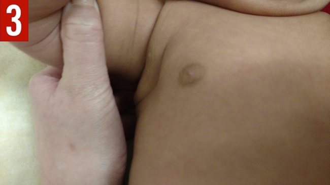
Based on the physical examination findings, the patient received a clinical diagnosis of polymastia (pseudomamma) in the axillary line.
Discussion. Breast development begins during the fifth week of gestation and originates from the mammary ridges, which run from the axillae to the inguinal region. In typical development, all but the region over the fourth intercostal space atrophies. In instances in which atrophy fails to occur, supernumerary breast tissue, or polymastia, results. Polymastia also less commonly may result from atypical ectoderm migration; in these cases, breast tissue is found outside the mammary ridge, such as on the thigh, shoulder, or back.
In 1915, Kajava developed the following 8 categories to describe polymastia, a system which is still in use today1:
- Class I, nipple, areola, and glandular breast tissue (polymastia)
- Class II, nipple and gland tissue with absence of areola
- Class III, areola and gland tissue with absence of nipple
- Class IV, glandular tissue alone
- Class V, areola and nipple with absence of gland tissue (pseudomamma)
- Class VI, nipple alone (polythelia)
- Class VII, areola alone (polythelia areolaris)
- Class VIII, patch of hair along the mammary ridge (polythelia pilosa)
Cases of polymastia are typically sporadic and asymptomatic. However, at the onset of puberty, the tissue may respond to the increased circulation of hormones, leading to growth of breast tissue. Surgery may be considered for cosmetic reasons and to avoid neoplasia.
Genitourinary anomalies have also been associated with the presence of polymastia, including an increased risk of duplicated excretory systems and polycystic kidney disease, as well as prostate, urinary, and renal cancers. Renal ultrasonography can be performed to assess for urinary tract abnormalities; if the results are positive, further imaging of the pelvis may be considered, given that the urogenital tracts develop together, and additional congenital abnormalities may be present.2,3
References:
- Patel PP, Ibrahim AM, Zhang J, Nguyen JT, Lin SJ, Lee BT. Accessory breast tissue. Eplasty. 2012;12:ic5.
- Ferrara P, Giorgio V, Vitelli O, et al. Polythelia: still a marker of urinary tract anomalies in children? Scand J Urol Nephrol. 2009;43(1):47-50.
- Varsano IB, Jaber L, Garty B-Z, Mukamel MM, Grünebaum M. 1984. Urinary tract abnormalities in children with supernumerary nipples. Pediatrics. 1984;73(1):103-105.