
Key words: early childhood caries, dental trauma, delayed primary dentition, orthodontic abnormalities, fluoride supplementation
A pediatric resident recommends an extensive evaluation for a 5-year-old girl who presents with a 1-week history of a neck mass and no other symptoms. According to the resident, physical examination findings are normal except for a 4 ×2-cm tender submandibular lymph node on the left. Before ordering laboratory and imaging studies, the attending pediatrician inspects the patient's teeth and finds multiple sites of dental decay and a deep, black cavitated lesion in a left mandibular molar. Severe dental decay is the likely cause of the lymphadenopathy. Antibiotics are prescribed, and the patient is referred to the pediatric dentist for definitive diagnosis and treatment.
Although the pediatrician cannot administer the definitive treatment for this child, an extensive lymphadenopathy evaluation was avoided by a simple look at the child's teeth.
This not-so-uncommon clinical scenario emphasizes the importance of inspecting the teeth as a part of the physical examination. It also emphasizes the need for pediatricians to understand common dental abnormalities that often present initially in the primary care setting.
Here we offer a review of early childhood caries (ECC), dental trauma, delayed dentition, and orthodontic abnormalities.
Often touted as the most common chronic disease of childhood, ECC is defined as decay in the primary teeth of children aged 71 months or younger.1 Surveillance during 1999 to 2002 in the United States found that 41% of children aged 2 to 11 years had dental caries in their primary teeth.2 Approximately one fifth of these children had untreated tooth decay.2 Nonhispanic white children and those from higher-income families had the lowest rate of caries.
Because of the frequency with which caries occur in American children, most pediatricians probably see patients with untreated dental decay on a weekly, if not daily basis. Physicians are obligated to recommend dental treatment for affected children and to help overcome any barriers to the receipt of care. If, in the pediatrician's opinion, the parent willfully fails to seek dental care for the child despite barrier removal, the parent may be responsible for dental neglect as defined by the American Academy of Pediatric Dentistry (AAPD).3 Dental neglect must be reported to Child Protective Services like any other form of abuse or neglect.
Pediatricians play an important role in preventing ECC. According to the AAPD and the American Academy of Pediatrics (AAP),4,5 caries prevention strategies that can be discussed at well-child visits include the promotion of daily flossing, toothbrushing with fluoridated toothpaste, and limited intake of sugar. Recommendations also include the establishment of a dental home--an approach to ensuring that all children have access to oral health education, oral health exams, and oral health treatments.6Ideally, soft drinks should be avoided completely. Juice should not be introduced into youngsters' diets before 6 months of age, nor should it be served in bottles or offered at bedtime.7 Advise parents of children younger than 3 years to clean the child's teeth as soon as the first tooth erupts and to avoid food-and utensil-sharing to decrease vertical transmission of Streptococcus mutans. The AAPD also promotes parental use of xylitol chewing gum. Evidence suggests that chewing 4 pieces of gum each day by the child's mother or caregiver significantly decreased the child's caries rates.5,8 Xylitol is a sugar substitute that inhibits growth of S mutans.
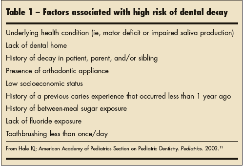
The AAPD also recommends the Caries-Risk Assessment Tool (CAT)9 to identify children at high risk for caries. The CAT is available on the AAPD Web site.9 Some factors listed in the CAT that signal increased risk of caries are cited in Table 1.
When inspecting a child's mouth, look for cavitated or noncavitated dental lesions, dental fillings, and missing teeth. The presence of gingivitis and/or plaque, chalky white spots, or deep fissures on the teeth are evidence of dental decay.9 Refer any child with worrisome findings to the pediatric dentist.
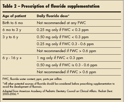
The pediatrician can have a great impact on ensuring that children obtain needed dental care. A systematic literature review found that children referred to a dentist by a primary care provider were more likely to visit a dentist than children who had not been referred.10 Along with the AAPD, the AAP supports the establishment of a dental home by 1 year of age, especially for those patients at high risk for ECC.11 Fluoride supplementation for children exposed to inadequate amounts in the water supply can be prescribed by the pediatrician as recommended by the AAPD (Table 2).12
| The Pathophysiology of Dental Caries: A Brief Review The cause of early childhood caries (ECC) is multifactorial, but the 2 main culprits that interact at the tooth surface to produce ECC are dietary sugar and bacteria—particularly Streptococcus mutans. Infants are known to acquire S mutans from their mothers or primary caregivers in the first 2 years of life.29,30 Exposure to sugary drinks from a bedtime bottle or frequent consumption of carbonated soft drinks31 places young children at increased risk for dental decay. The acidic by-products of the bacterial metabolism of sugary substances lead to the development of plaque. Plaque adheres to the tooth surface and causes dental demineralization or the leaching out of important minerals (ie, calcium, phosphate, and carbonate) (A). 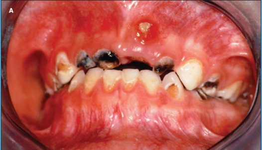 Untreated dental caries can lead to periodontal abscesses (B). Because of the adjacent location of anatomic structures, facial cellulitis (C) and maxillary sinusitis may result from direct extension of the odontogenic infection, which causes tissue breakdown and bone resorption.32 Further extension of this infectious process could potentially affect intracranial structures, leading to brain abscess or orbital cellulitis. Untreated odontogenic infections can also lead to Ludwig angina—a rare severe condition involving extension of infection into the floor of the mouth and beyond, with potential airway compromise.33 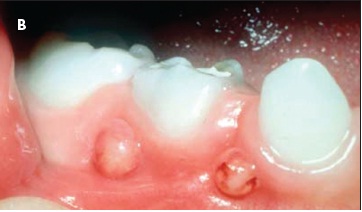 As with all odontogenic infections, the most common causative agents are anaerobes and Streptococcus and Staphylococcus species. A patient with signs and symptoms of Ludwig angina (neck and/or mouth pain and swelling, trismus, fever, malaise, fetid breath) must be treated emergently to ensure a secure airway. Surgical debridement as well as administration of broad-spectrum antibiotics with adequate anaerobic coverage provide definitive treatment.33 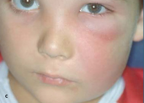 |
Dental Trauma
Although dentists are obligated to be available for emergent dental care,13 the general pediatrician or acute care physician is often the first one called or visited after dental trauma occurs. Dental trauma can lead to tooth fractures and displacements of varying degrees, intrusions (apical displacement of tooth into the alveolar bone), extrusions (partial displacement of tooth axially from the socket), and avulsions (complete displacement of a tooth from its socket). If severe trauma has occurred or is suspected, immediately refer the patient to an emergency facility where dental and other medical concerns can be addressed simultaneously.
Rowley and colleagues14 described over 1000 patients who presented to an emergency medical facility after dental trauma. Most trauma occurred in children 1 to 3 years of age. After age 8, boys experienced more dental trauma than girls. The maxillary incisors were most commonly affected. In older children and adolescents, sports-related oral injuries are a frequent cause of dental trauma.15
A child with a dental injury should be approached like any other patient to ensure that all medical issues are addressed. The history and physical examination determine treatment. If the patient has sustained head trauma, pay particular attention to neurological function. Also, determine whether the patient is adequately immunized against tetanus.16 Worrisome dental signs and symptoms include tooth pain (spontaneous or secondary to touch or pressure), tooth temperature sensitivity, increased tooth mobility and/or obvious displacement, or discoloration of the tooth.16 Intrusion causes affected teeth to have a shortened appearance; extrusion causes a lengthened appearance.
The management of dental trauma is almost always best left to a dentist. Andreasen and colleagues17 provide suggested time frames in which children with certain dental injuries need treatment by a dental expert. In general, dental injuries should be treated acutely (within a few hours) or subacutely (within 24 hours). Some crown and crown/root fractures may fare well with a delayed approach (after the first 24 hours),17 but that decision should rest with the dental expert.
When the pediatrician is contacted about a child with dental intrusion (which often follows an accident, such as a face-first fall down a playground slide), the goal is to expedite referral to the dentist who will determine (with radiographs) whether the permanent teeth are affected. If the permanent teeth are not affected and the primary teeth are otherwise asymptomatic, reassurance along with allowing the spontaneous re-eruption of teeth is the usual treatment.
Avulsion of a permanent tooth is an emergency. Parents and coaches need to be aware that reimplantation of the avulsed tooth back into its own socket within 5 minutes offers the best prognosis.13 Debris can be gently rinsed off the tooth if it is reinserted. If reimplantation on the spot is not feasible, the tooth should be stored in a medium (in order of preference: commercial dental solutions, cold milk, saliva, saline, or water) while the patient is en route to the dentist.13 According to the AAPD,13 teeth should not be reimplanted in patients who have immune deficiencies, severe congenital cardiac anomalies, severe uncontrolled seizures, mental disabilities, uncontrolled diabetes, or lack of alveolar integrity.
During well-child visits, counsel patients about the importance of using protective mouth guards and face protectors during sports activities.13,18,19 The dental literature supports the efficacy of properly fitted mouth guards in reducing sports- related oral injuries.19 The importance of immediate reimplantation of an avulsed tooth is a helpful pointer for parents and coaches.
Deciduous teeth erupt in a predictable pattern beginning at age 5 to 7 months and ending at age 2 to 3 years. Children who lack teeth by their first birthday cause much parental concern. If no other abnormalities are present, however, delayed primary dentition is probably not associated with any pathology. A watch-and-wait approach is appropriate, along with co-monitoring in the dental home, which should be established by age 1. Ethnic and nutritional status can affect the timing of tooth eruption. For example, Al-Jasser and Bello20 found that Saudi children experience a slightly delayed primary tooth eruption compared with the norms. Holman and Yamaguchi21 report delayed emergence of primary teeth in poorly nourished Japanese children.
Rarely, late eruption of primary teeth is the result of an underlying systemic condition, such as genetic syndromes, endocrine disorders (ie, hypothyroidism, hypoparathyroidism, hypopituitarism), complications of medical interventions (chemotherapy, radiation) and nutritional deficiencies (ie, rickets). At least 50 genetic disorders are associated with delayed tooth eruption.22 Prematurity,23 increasing birth number (ie, young children with a number of older siblings), and infection with HIV have also been implicated.24
The evaluation of the child who lacks teeth by age 1 year includes a review of the medical and family history and a thorough physical examination for evidence of an underlying systemic illness. A history of prematurity, poor nutrition, and any systemic symptoms should be noted. The physical examination focuses on the presence of oral pathology, dysmorphic features, bony abnormalities, growth disturbances, and other findings that indicate an endocrinopathy or genetic disorder.
Inspect the child's mouth for any soft tissue pathology. Carefully palpate the alveolar ridges to ensure the absence of any budding teeth. Laboratory studies are obtained on a case-by-case basis and are guided by the suspected underlying systemic illness (eg, tests of thyroid-stimulating hormone and thyroxine levels in suspected hypothyroidism).
The workup for delayed dentition by the pediatric dentist usually includes a panoramic radiograph for evaluation of any oral pathology and assessment of teeth development. Treatment consists of observation and diagnosis and comprehensive management of any underlying conditions. The rare condition of congenital absence of teeth may require the expertise of other oral health professionals, such as the orthodontist and oral surgeon.
Orthodontic abnormalities arise from malaligned or misplaced teeth, too many or too few teeth, crowded teeth and jaw, and facial growth discordance.Premature loss of the primary teeth secondary to dental caries can also lead to orthodontic abnormalities. Common abnormal occlusal characteristics in the US population include diastema (a gap between 2 teeth), malalignment of the lower and upper anterior teeth, posterior crossbite, and overbite. Data from the third National Health and Nutrition Examination Survey25 revealed that one fifth of Americans have bite abnormalities; of these, 2% are disfiguring. Brunelle and coworkers26 report that 8% of the US population has a severe overbite of 6 mm or more. Orthodontic abnormalities are a source of both cosmetic concern and oral dysfunction.
Inheritance plays a large role in the development of malocclusion. Many genetic syndromes (ie, ectodermal dysplasia) are accompanied by dental abnormalities. Acquired orthodontic abnormalities are the result of prolonged digit or pacifier sucking habits, obligate mouth breathing, dental disease or trauma, dysfunctional swallowing, and malnutrition.
The child with a mild orthodontic abnormality may not have any complaints. However, a severe orthodontic abnormality may adversely affect self-esteem, cause pain and masticatory dysfunction, wear down teeth surfaces prematurely, and increase the risk of caries because of difficult-to-clean areas. Inspection of the mouth will reveal obvious displacements of teeth, but subtle findings may be missed. The need for the dental expert's input cannot be overstated.
The American Association of Orthodontists27 recommends that all children should be examined by an orthodontist at age 7 years. By age 7, the first permanent molars have erupted and the posterior occlusion is established, incisors have begun to erupt, and dental crowding and bite and jaw disturbances can be detected. It is reasonable to refer a child of any age with a suspected orthodontic abnormality--especially if the acute and long-term consequences are unknown.
Treatment of children with orthodontic abnormalities is tailored to the patient's particular clinical circumstance. Although people in all walks of life can suffer from occlusal disorders, there is a social discrepancy in the frequency of treatment for orthodontic conditions; those with higher incomes are much more likely to obtain treatment.25 The exorbitant cost of some orthodontic interventions is a great barrier to receiving care. Braces are quite common and can cost a few thousand dollars, while more complicated procedures involving implants and prosthetics for a patient with ectodermal dysplasia can cost up to $40,000.28 Strategies to obtain orthodontic care for lower income children are needed and represent a whole topic unto itself.
REFERENCES:
1. American Academy of Pediatric Dentistry. Definition of early childhood caries (ECC). Reference Manual 2005-2006. Adopted 2003. Available at: http://www.aapd.org/media/Policies_Guidelines/D_ECC.pdf. Accessed on June 25, 2007.
2. Beltrán-Aguilar ED, Barker LK, Canto MT, et al. Surveillance for dental caries, dental sealants, tooth retention, edentulism, and enamel fluorosis—United States, 1998-1994 and 1999-2002. MMWR. 2005;54:1-43.
3. American Academy of Pediatric Dentistry. Definition of dental neglect. Pediatr Dent. 1997;19:24.
4. American Academy of Pediatric Dentistry. Guideline on infant oral health care. Clinical Guidelines. Adopted 1986. Last revised 2004. Available at: http://www.aapd.org/media/Policies_Guidelines/G_InfantOralHealthCare.pdf. Accessed June 25, 2007.
5. Oral Health Risk Assessment Timing and Establishment of a Dental Home. Pediatrics. 2003;111:1113-1116. Available at: http://aappolicy.aappublications.org/cgi/content/full/pediatrics;111/5/1113. Accessed June 25, 2007.
6. Oral Health Risk Assessment. American Academy of Pediatrics. Available at: www.aap.org/commpeds/dochs/oralhealth/ohra/Dental%20Home%20Model.pdf. Accessed July 7, 2007.
7. Committee on Nutrition. American Academy of Pediatrics: The use and misuse of fruit juice in pediatrics. Pediatrics. 2001;107:1210-1213.
8. Isokangas P, Soderling E, Pienihakkinen K, Alanen P. Occurrence of dental decay in children after maternal consumption of xylitol chewing gum, a follow-up from 0 to 5 years of age. J Dent Res. 2000;79:1885-1889.
9. American Academy of Pediatric Dentistry. Policy on the use of a caries-risk assessment tool (CAT) for infants, children, and adolescents. Adopted 2002, revised 2006. Available at: http://www.aapd.org/media/Policies_Guidelines/P_CariesRiskAssess.pdf. Accessed June 25, 2007.
10. Bader JD, Rozier RG, Lohr KN, Frame PS. Physicians’ roles in preventing dental caries in preschool children—a summary of the evidence for the US Preventive Services Task Force. Am J Prev Med. 2004;26;315-325.
11. Hale KJ; American Academy of Pediatrics Section on Pediatric Dentistry. Oral health risk assessment timing and establishment of the dental home. Pediatrics. 2003;111(5, pt 1):1113-1116.
12. American Academy of Pediatric Dentistry. Guideline on fluoride therapy. Reference Manual 2005-2006. Available at: http://aapd.org/media/policies_guidelines/g_fluoridetherapy.pdf. Accessed June 25, 2007.
13. American Academy of Pediatric Dentistry Council on Clinical Affairs. Guideline on management of acute dental trauma. Pediatr Dent. 2005-2006;27:135-142.
14. Rowley ST, Sheller B, Williams BJ, Mancl L. Utilization of a hospital for treatment of pediatric dental emergencies. Pediatr Dent. 2006;28:10-17.
15. Cornwell H. Dental trauma due to sport in the pediatric patient. J Calif Dent Assoc. 2005;33:457-461.
16. Bernius M, Perlin D. Pediatric ear, nose and throat emergencies. Pediatr Clin North Am. 2006;53:195-214.
17. Andreasen JO, Andreasen FM, Skeie A, et al. Effect of treatment delay upon pulp and periodontal healing of traumatic dental injuries—a review article. Dent Traumatol. 2002;18:116-128.
18. American Academy of Pediatric Dentistry Clinical Affairs Committee; American Academy of Pediatric Dentistry Council on Clinical Affairs. Policy on prevention of sports-related orofacial injuries. Pediatr Dent. 2005-2006;27(7 Reference Manual):45.
19. ADA Council on Access, Prevention and Interprofessional Relations; ADA Council on Scientific Affairs. Using mouthguards to reduce the incidence and severity of sports-related oral injuries. J Am Dent Assoc. 2006;137:1712-1720.
20. Al-Jasser NM, Bello LL. Time of eruption of primary teeth in Saudi children. J Contemp Dent Prac. 2003;4:1-7.
21. Holman DJ, Yamaguchi K. Longitudinal analysis of deciduous teeth emergence, IV: covariate effects in Japanese children. Am J Phys Anthropol. 2005;126:352-358.
22. Suri L, Gagari E, Vastardis H. Delayed tooth eruption: pathogenesis, diagnosis, and treatment. A literature review. Am J Orthod Dentofacial Orthop. 2004;126:432-445.
23. Viscardi RM, Romberg E, Abrams RG. Delayed primary tooth eruption in premature infants: relationship to neonatal factors. Pediatr Dent. 1994;16:23-28.
24. Noffke CE, Chabikuli NJ, Nzima N. Impaired tooth eruption: a review. SADJ. 2005;60:422-425.
25. Proffit WR, Fields HW, Moray LJ. Prevalence of malocclusion and orthodontic treatment need in the United States: estimates from NHANES III survey. Int J Adult Orthodon Orthognath Surg. 1998;13:97-106.
26. Brunelle JA, Bhat M, Lipton JA. Prevalence and distribution of selected occlusal characteristics in the US population, 1988-1991. J Dent Res. 1996;75:706-713.
27. American Association of Orthodontists. American Association of Orthodontists Recommendation for Orthodontic Check-Ups No Later Than Age 7. Available at: http://www.braces.org/healthcareprofessionals/dentists/upload/Prolems_to_Watch_for_in_7YrOlds_FINAL.pdf. Accessed June 26, 2007.
28. Murdock S, Lee JY, Guckes A, Wright JT. A cost analysis of dental treatment for ectodermal dysplasia. J Am Dent Assoc. 2005;136:1273-1276.
29. Wan AK, Seow WK, Purdie DM, et al. A longitudinal study of Streptococcus mutans colonization in children after tooth eruption. J Dent Res. 2003;82:504-508.
30. Li Y, Caufield PW. The fidelity of initial acquisition of mutans streptococci by infants from their mothers. J Dent Res. 1995;74:681-685.
31. Sohn W, Burt BA, Sowers MR. Carbonated soft drinks and dental caries in the primary dentition. J Dent Res. 2006;85:262-266.
32. Mehra P, Murad H. Maxillary sinus disease of odontogenic origin. Otolaryngol Clin North Am. 2004;37:347-364.
33. Belleza WG, Kallman S. Otolaryngologic emergencies in the outpatient setting. Med Clin North Am. 2006;90:329-353.